

COVID19 Vaccines and Altered Vascular Rheology.
Spike associated destruction of blood vessels is a central pathological mechanism of CoVax Disease. Obstruction to blood flow from clots and or bleeding from blood vessel damage is collateral damage.


Flow of molten rock and gas from the eruption of Kilauea on the Big Island, Hawaii. Reprinted with Permission of Lewis Photography
Rheology is the study of the flow of matter, primarily in a fluid (liquid or gas) state but also as "soft solids" or solids under conditions in which they respond with plastic flow rather than deforming elastically in response to an applied force.[1] Rheology is the branch of physics that deals with the deformation and flow of materials, both solids and liquids.-Wikipedia
Rheology has broader application than does the term “circulation” which is an oversimplification of what actually happens in the human body in terms of the numerous types of material flow.
The concept applies to the nourishing of hyaline cartilage in a joint by synovial fluid. The knee, for example, has an elaborate mechanism to circulate a tiny amount of nourishing and lubricating synovial fluid throughout the joint. The medial and lateral menisci are part of this mechanism.
Additional examples are the flow of lymphatic fluid throughout the body and cerebrospinal fluid in the brain and spinal cord.
This article will explore novel pathology affecting vascular rheology following Covid19 “vaccines”.
Burkhardt/Lang Case 39b Presentation: 42 y/o MD
History
Case 39b is that of a 42 year old physician who presented to a vascular clinic with multiple physical complaints after a bout of COVID19 in January of 2021 followed by a single dose of Comirnaty (BNT162b2) on July 8, 2021.
Her complaints consisted of general fatigue, reduced resilience, and a cognitive disorder. Her vascular symptoms consisted of vasospasms and microcirculatory disorders in her feet. Her toes would turn white and feel numb. She was symptomatic at rest as well as with activity.
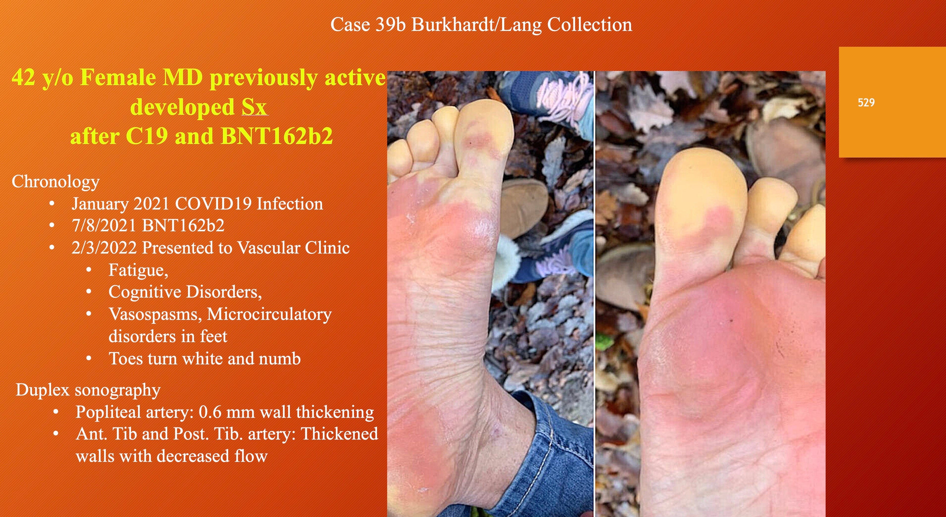
Findings:
Inspection of her foot revealed reddish discoloration of the plantar skin and pale toes.
Ultrasound examination of the arterial system identified thickening of the popliteal, anterior and posterior tibial arteries in her right foot. The status of the third vessel, the peroneal or fibular artery is less accessible and its status was not reported.
The popliteal artery takes off from the superficial femoral artery in the lower thigh and extends to the branching of the anterior tibial artery below the knee. The tibial arteries are typically sampled at the level of the ankle.
The implication is that the arterial wall is thickened over an extensive zone from thigh to foot. The question remaining is how extensive is the vascular pathology, what other vessels have such change.

Histopathological and Immunohistochemical Findings

Biopsies of skin revealed what was described as obliterating endothelialitis (inflammation of the cells lining the lumen of arteries) with lymphocytic infiltration of smaller arteries (center above) and some some disruption of the muscular layer of larger arteries.

This vessel (above) has a thickened wall that stains positive with Congo Red stain for amyloid-like proteins accounting for decreased arterial compliance reduced blood flow.

This photomicrograph above shows spike protein in a capillary. Spike stains were positive. Nucleocapsid stains were negative. This combination implicates the corona vaccine and not the virus.

Skin biopsy showing perivascular lymphocyte infiltration and obliteration of the vascular lumen (left). Spike protein is seen in the vessel wall (right).
The conclusion from review by Drs. Burkhardt and Lang is that the vascular thickening and histological change in the biopsy specimens was caused by the “vaccine”.
Diagnosis: Pronounced, partially obliterating inflammatory vasculopathy with amyloid deposits and positive expression of the spike protein. Superficial periadnexal dermatitis with minor predominantly lymphocytic panniculitis. Proliferation of interepithelial Langerhans cells; focal with spike protein expression in the epidermis.
The amyloid deposits in the vessel walls and the positive findings for the spike protein suggest an association and probable causative role of the coronavirus-associated and vaccine-associated spike protein.
The perivascular, almost exclusively lymphocytic vasculitis and the marked proliferation of dendritic Langerhans cells in the epidermis indicate an ongoing immunologic-inflammatory process.- Burkhardt and Lang, Case 39b.
Thickened, inflamed arteries in the popliteal-tibial system as well as smaller vessels explain this person’s symptoms both in her leg and foot but also her cognitive disorder as a similar process is likely going on in her central nervous system.
This case was made more complex by the discovery of a cold activated white fibrous clot, now referred to as a Calamari Clot. Dr. McCairn has studied Calamari clots submitted by embalmer Richard Hirschman.

Cryoproteins/Cryoglobulins
Dr. Burkhardt took a serum specimen from the patient and refrigerated it overnight. A whitish, rubbery clot formed under refrigeration implying the presence of cold activated proteins called cryoproteins. Whitish clots had not been identified in the biopsy specimens possibly because her large vessels were not thoroughly examined in her popliteal/tibial arteries.


The white substance in the image above is the clot that formed overnight. Cold activation is a feature of these so called Calamari clots since they were identified after refrigeration but they also form at normal human body temperature. The name calamari describes the rubbery consistency of these novel clots. (Specimen below courtesy of Richard Hirschman).

Under low magnification the white clot material has a fibril structure (below) that Dr. McCairn compared with mycelia of fungi in appearance but with no connection to actual fungi.
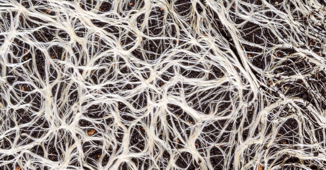
Above, Fungal mycelia.

Here is a magnified image showing interwoven fibrils with increased magnification in the image below.

Lymphocytes were also identified, purple-blue dots in the image above.

Periodic Acid-Schiff (PAS) stain was positive for glycoproteins.

Elastic von Gieson stain for elastin fibers was positive.
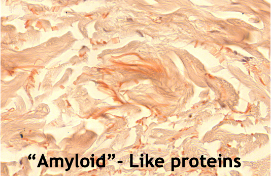
Amyloid-like proteins were identified by Congo Red stain (above).
Further analyses of the protein structure using Rahman spectroscopy and other techniques were performed to further characterize the proteins in the clot.

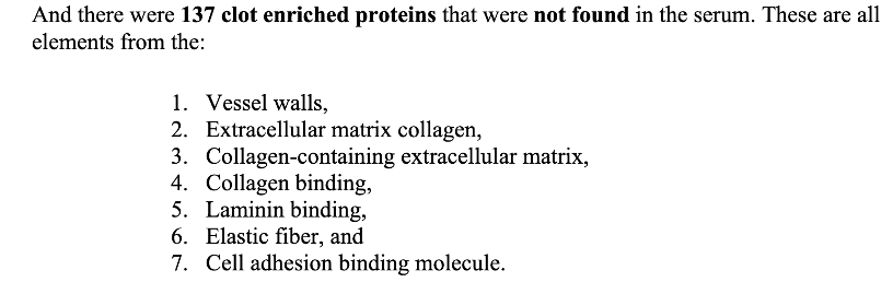
The origin of these proteins has not been precisely determined but one possibility is that the widespread microangiopathy causes debris from the areas of dense lymphocytic infiltration with discharge of the “biosoup” into the circulation is a contributor.
Case 17 below illustrates the possible mechanism as the arterial wall is degraded by activated lymphocytes, a complex brew is released into circulation causing stasis and clotting. This person was found dead, probably from a stroke.

Higher power magnification shows what appears to be release of the cellular and molecular debris into the arterial lumen.
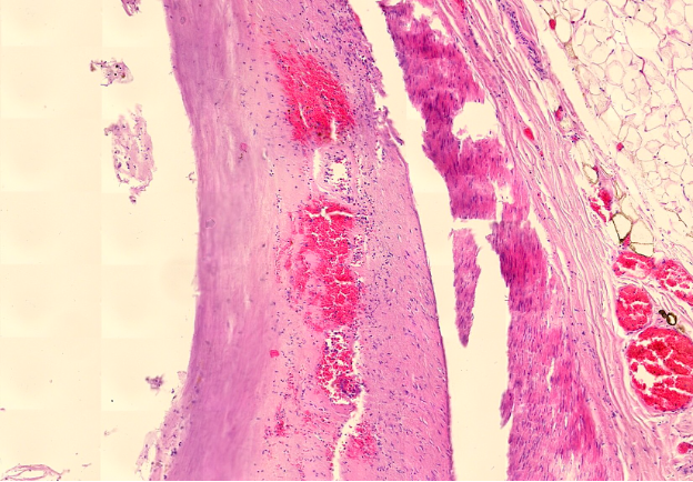
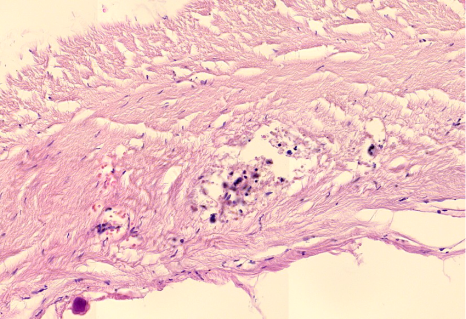
The above specimen from an autopsy shows the physical deterioration of the muscular layer of a large artery.
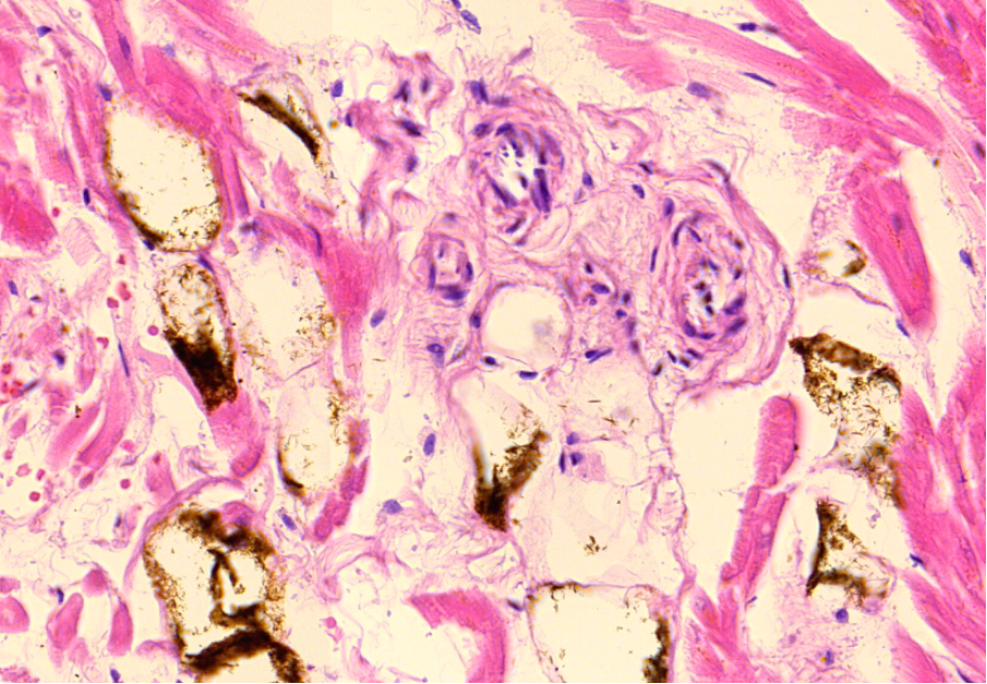
Heart specimen from another autopsy shows degenerating muscle tissue with odd brown-black inclusions of unknown composition and origin.

Dense lymphocytic infiltration and black inclusions with destruction of tissues.
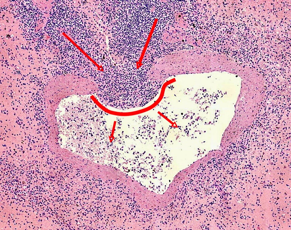
The specimen above from yet another subject shows obliteration of what was a probably a vein that is now filled with spindle like cells.
Conclusion:
Case 39b is that of a young physician with evidence of large and small vessel disease probably caused by BNT162b2. The large vessel in this instance is the popliteal artery and its tributaries, the anterior and posterior tibial arteries.
No arteriogram was reported so it is not known how extensive this woman’s vascular problem is and what the options are for angioplasty and stent, bypass or interposition graft if limb viability becomes threatened by progression of the inflammatory process. Bypass is not effective for ischemia caused by small vessel disease.
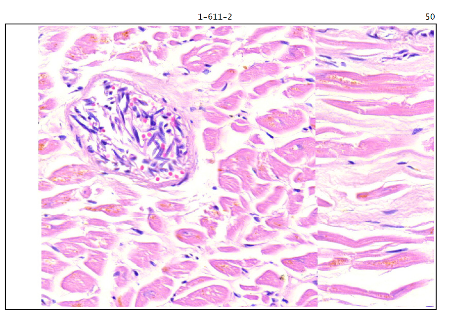
Her CNS symptoms could be related to vaccine induced microangiopathy as in Case 44 above showing destruction of the muscle layer of an artery in the dura mater, a thick membrane covering the brain.
It was not determined that the Calamari Clot phenomena was active in this woman as the strange white fibrous clot was identified in a centrifuged and refrigerated serum specimen and not the living person. Neither was it “ruled out”.
Is the white fibrous material from the refrigerated specimen in Burkhardt and Lang Case 39b the same as what was seen by Dr. McCairn? Possibly. Here is what AI (Grok3) came up with,
Amyloid accumulation is a problem as it compromises function of the tissues it is deposited in.
Amyloids are aggregates of proteins characterized by a fibrillar morphology of typically 7–13 nm in diameter, a β-sheet secondary structure (known as cross-β) and ability to be stained by particular dyes, such as Congo red.[1] In the human body, amyloids have been linked to the development of various diseases.[2] Pathogenic amyloids form when previously healthy proteins lose their normal structure and physiological functions (misfolding) and form fibrous deposits within and around cells. These protein misfolding and deposition processes disrupt the healthy function of tissues and organs.-Wikipedia
The McCairn analysis not only confirmed the presence of amyloid, but of prion type amyloid.
The term prion comes from "proteinaceous infectious particle". Unlike other infectious agents such as viruses, bacteria, and fungi, prions do not contain nucleic acids (DNA or RNA). Prions are mainly twisted isoforms of the major prion protein (PrP), a naturally occurring protein with an uncertain function. They are the hypothesized cause of various TSEs, including scrapie in sheep, chronic wasting disease (CWD) in deer, bovine spongiform encephalopathy (BSE) in cattle (mad cow disease), and Creutzfeldt–Jakob disease (CJD) in humans.-Wikipedia
Let us hope the prion issue goes away.
However, three cases of Creutzfeld Jakob Disease were reported in Hood River County, Oregon.

Grok3 was asked to compute the probability of 3 cases of CJD in Hood River County, Oregon.
The statistical probability of observing three or more cases of Creutzfeldt-Jakob disease in Hood River County (population ~24,000) over eight months, given an incidence rate of 1.5 cases per million per year, is approximately 0.0019% (or 1 in 52,632). This indicates that the occurrence of three cases is a highly unusual event, statistically speaking, though it does not necessarily imply a common cause or public health risk.
Hopefully our new public health officials will give this the attention it deserves.
Thank you
Thank You for this post. Just one remark, your username 'Experimental Gene Therapy' is NOT 'in phase' with the title of this post:"COVID19 Vaccines and Altered Vascular Rheology." It should be 'Covid19 EXPERIMENTAL GENE Therapies and altered Vascular Rheology'...
'